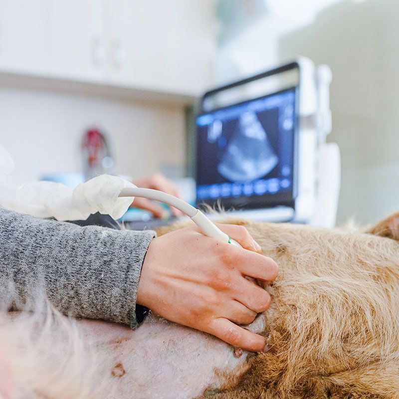
Ultrasound uses sound waves to create real-time images of internal structures. Unlike X-rays, which show bones and air-filled spaces, ultrasound excels at visualizing soft tissues and fluid-filled areas. We use this technology to examine the liver, kidneys, spleen, bladder, intestines, and other abdominal organs. We can also evaluate the heart to check for structural problems or fluid accumulation.
This imaging method is completely safe and painless. Most pets tolerate ultrasound examinations well, though we may need to shave a small area of fur to improve contact between the ultrasound probe and skin. The procedure typically takes 20 to 40 minutes, depending on which structures we’re examining and what we find.